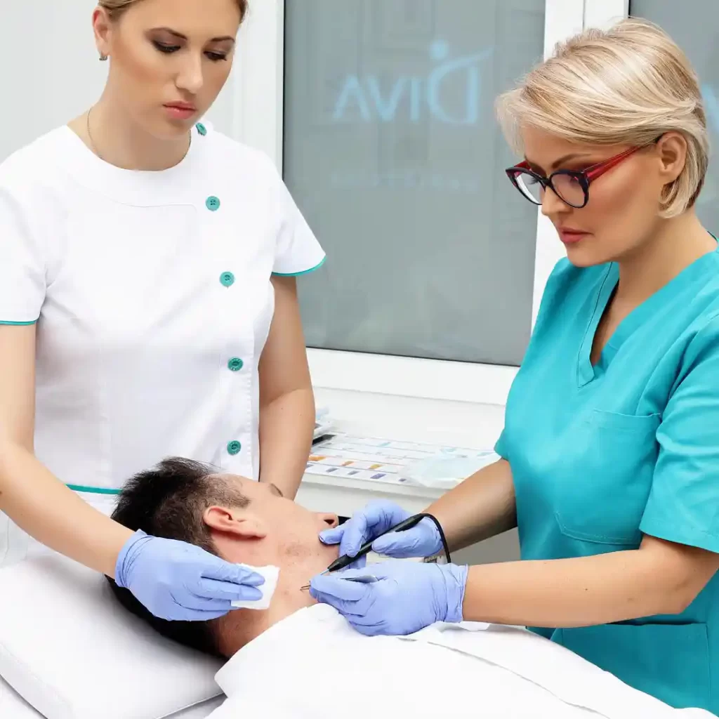
Free consultation before the procedure during which radio wave surgery is used at Diva Polyclinic
You can schedule a FREE consultation for the removal of moles, warts and other skin changes with the doctors of the Diva Dr. Bogdanović polyclinic in one of our polyclinics or specialist dermatology offices:
Radio waves have a high frequency of 3.8 MHz, which minimally traumatizes the tissue, so they can successfully replace the scalpel in all surgical branches.
The radio-wave technique is most widely used in plastic surgery and dermatosurgery, because it is precisely in these branches of medicine that it is most important to obtain the finest and almost imperceptible scar.
What can be removed with radio wave surgery?
- Moles – usually benign growths, but they should be monitored regularly because if they show changes, their preventive removal is necessary.
- Viral warts are skin changes caused by different strains of the HPV virus.
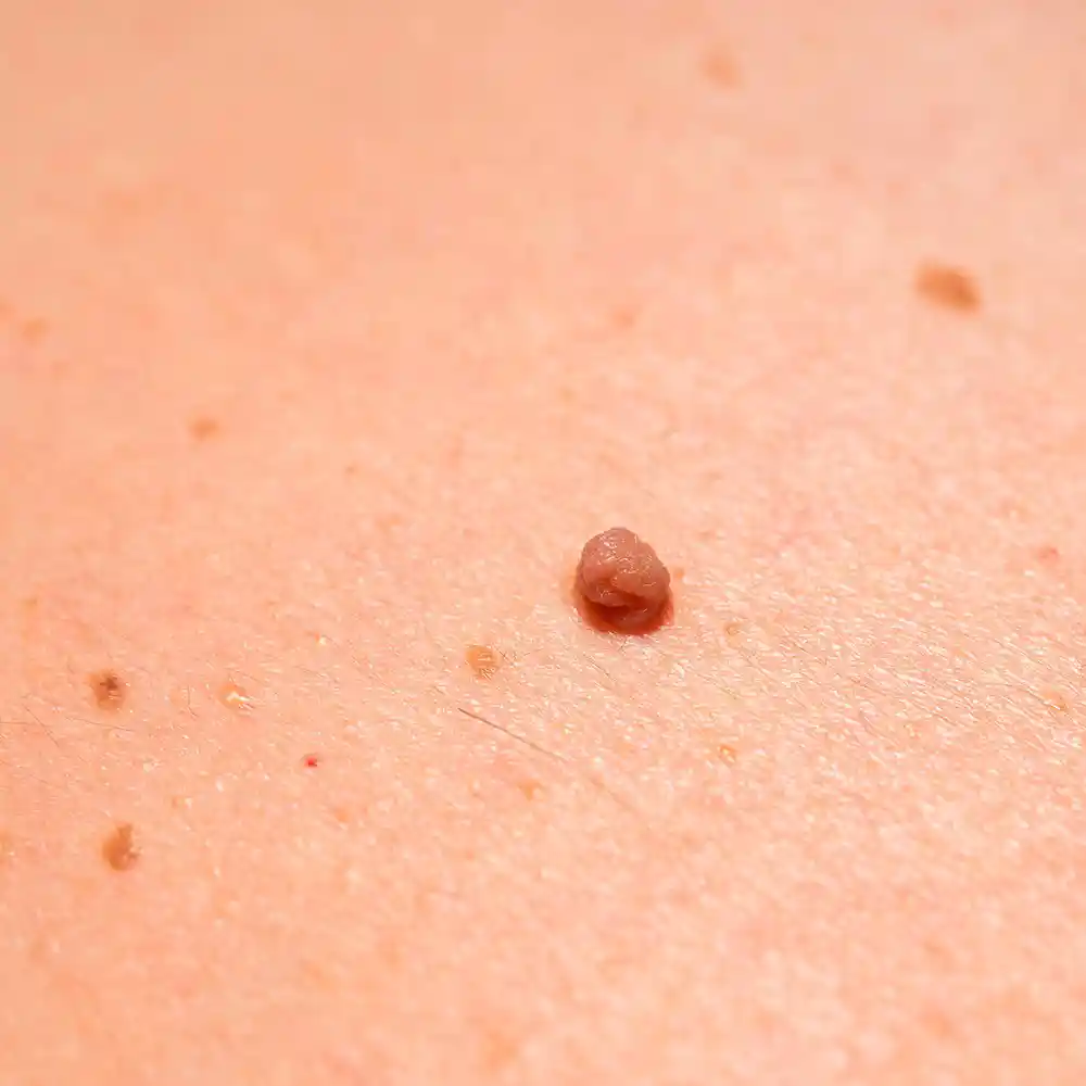
- Papillomas – hanging warts of viral origin.
- Fibromas – frequent growths on the skin of benign origin. They can be of different sizes and are usually on a stalk.
- Keratoses – are formed from seborrheic keratin deposits.
- Angiomas – often called red moles are benign changes of vascular origin.
- Sebaceous cysts (atheroma) are cysts that are the result of blockage of the sebaceous glands’ outlet channels.
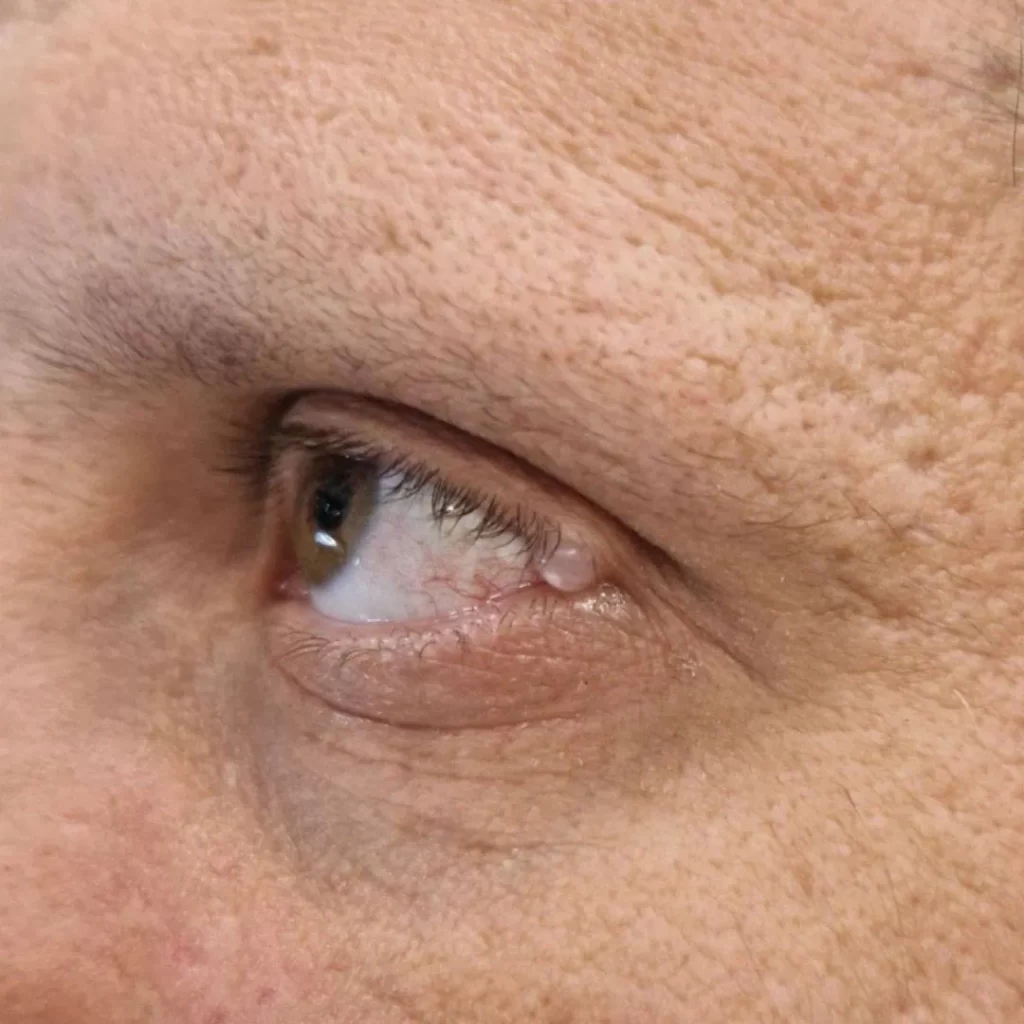
- Syringomas – small watery cysts that occur as a result of blockage of sweat glands and most often appear in the region of the eyelids.
- Xenthelasma – yellowish deposits that appear on the eyelids.
- Condylomas – genital warts caused by HPV viruses.
- Granulomas – changes that appear after a skin injury and tend to grow rapidly and tend to bleed.
- Dermatofibromas – growths on the skin that most often appear on the hands and feet.
Why is radiosurgery an excellent choice for removing moles, warts and skin growths?
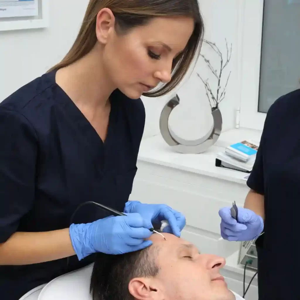
- comfortable (during the procedure a local anesthetic in the form of a cream is used),
- it takes a short time, ONLY 20 to 30 seconds per change,
- safe and
- so sophisticated that patients do not feel that they are undergoing surgical intervention,
which is important for stress reduction and the patient’s sense of comfort.
And in addition, the advantages of radio waves are minimal trauma to the surrounding tissue, which is crucial for:
- easier,
- better and
- faster wound healing,
- and the result is almost imperceptible scars (whether they will be noticeable depends on several factors such as genetics and the tendency of the skin to create them, as well as the age of the patient).
Radio waves have a high frequency of 3.8 MHz, which minimally traumatizes the surrounding tissue, resulting in a sophisticated precise cut, so that they can successfully replace the scalpel in almost all surgical branches! This technique is widely used in plastic surgery and dermatosurgery, because it is precisely in these branches of medicine that the maximum precision of the cut is important in order to obtain the finest and almost imperceptible scar.
All changes, except for viral warts, which were removed from the skin at the “Diva” polyclinic, MUST be sent for a histopathological examination, so that we carry out a triple control of the diagnosis:
- clinical examination by a dermatologist or plastic surgeon,
- dermoscopic examination and
- histopathological examination by a pathologist with the aim of maximum patient safety.

Advice from a specialist doctor regarding skin growths:
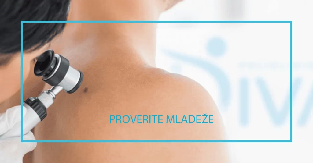
It is very important to make a timely diagnosis and carry out treatment. That is why it is necessary to regularly control all moles and other skin changes!
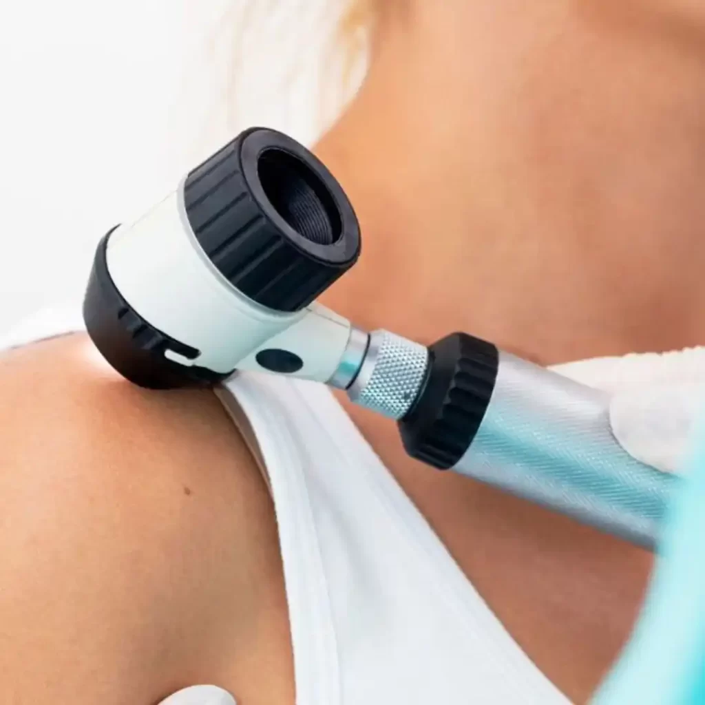
Examination of growths on the skin before intervention with radio waves - dermoscopy
Given that warts, moles and other growths on the skin can be potential foci of malignant tumors, preoperative diagnosis is extremely important. In addition to the clinical examination, it is sometimes necessary to perform a dermoscopic examination to make the diagnosis precise, which is crucial for excluding malignancy.
Pathohistological examination after radio wave surgery
Pathohistological examination means examination of the change that was removed by the radio wave technique under a microscope by a pathologist.
All changes, except for viral warts, which were removed from the skin at the “Diva” polyclinic, MUST be sent for histopathological examination.
What is radio wave surgery for?
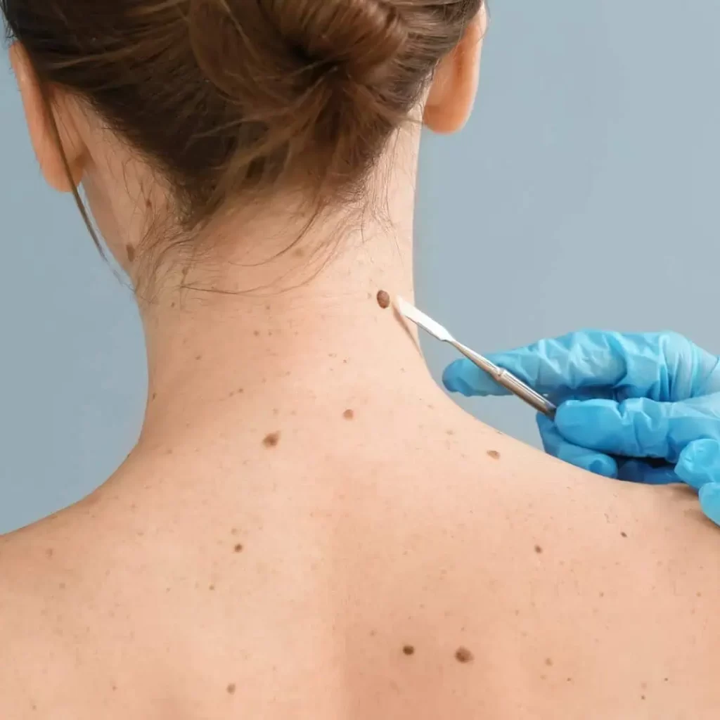
Removal of moles
Moles, naevus, are benign growths on the skin. These changes are a combination of accumulation of pigment cells and connective tissue. The color of moles can be from light to dark brown. Some appear already at birth, in a small percentage. And they can appear throughout life on all skin regions. They can change shape, color and size over time, so they need to be monitored regularly. However, more than 90% of moles do not belong to the risk group and can be treated for aesthetic or practical reasons, so radiowave dermatosurgical technique is optimal for their removal.
Removal of ALL types of warts and keratoses
Warts – verrucae, are skin changes of viral origin. This disease affects about 10% of the entire population. They most often appear on the hands, face, neck, under the armpits, and can also be on the back or stomach. Given that they are of viral origin, they spread from one part to another, say from the neck to the face or in the armpit region, and they can also be transmitted to people in the environment, so removing warts is recommended to prevent their spread. Several months can pass from incubation to the appearance of warts on the skin.
Types of warts that can be removed by radiosurgery
- ordinary – can appear on all regions of the skin
- verrucae plane – affect the face and neck,
- ridges – present on the neck and body,
- mollusks – they are characteristic for children’s age
- mosaic – most often appear on the hands and feet
- genital (condyloma) – transmitted sexually.
Removal of keratoses
Removal of atheroma, fibroma and xanthelasma
- lid,
- door,
- troop,
- armpit and
- groin.
Removal of syringoma, granuloma and dermatofibroma
Radio wave surgery - price
NOTE:
Whether radio waves are an adequate method for removing changes on the skin will be evaluated by a specialist doctor during a free consultation.
The results of the procedure may vary from patient to patient, and this is influenced by factors such as skin type, i.e. whether it is prone to scar tissue formation.
You can read the entire disclaimer HERE.
Appointment for a free examination before the procedure during which radio wave surgery is used in Belgrade
At Diva Dr. Bogdanović Polyclinic Belgrade, with our doctors specializing in dermatology and aesthetic surgery, you can schedule a FREE consultation for a pre-procedure examination during which radiowave surgery is used in one of our polyclinics or specialist dermatology offices:
to one of the phone numbers 063 338 334 or 011 3242 841 or to the email info@divaclinic.com
You can schedule an examination or procedure
· every working day from 9 a.m. to 8 p.m
· and on Saturdays from 9 a.m. to 3 p.m.
Price for specialist dermatological skin examination and dermoscopic examination of moles
If a specialist dermatological examination of the skin is required due to dermatological changes on the skin, then the price is RSD 6,000.
If you want to schedule only a dermoscopic mole examination, the price is RSD 6,000.
Your “Polyclinic Diva”
Author of the text: Dr. spec. Svetlana Bogdanović
Koautor: dr spec. Marija Šejnjanović